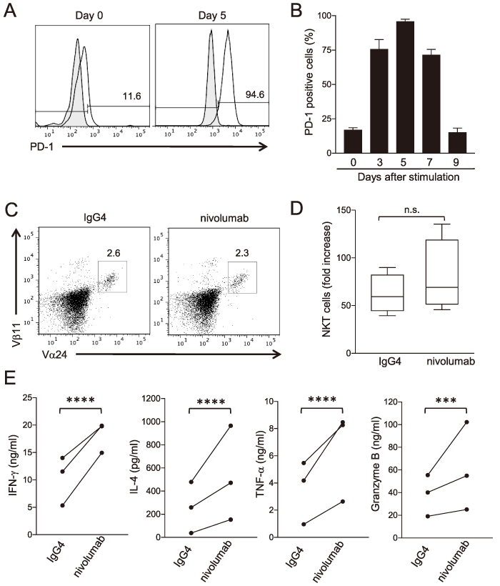
Fig. 1 Nivolumab enhances the production of cytokines and effector molecules by NKT cells. (A, B) PD-1 expression on NKT cells. PBMCs were cultured with α-GalCer and IL-2. Cells were harvested at days 0, 3, 5, 7, and 9, stained with an anti-PD-1 antibody, and analyzed by flow cytometry. (B) Bars and error bars indicate the mean and SD of technical triplicate, respectively (n = 3) . The data are representative of the results from two independent experiments. (C, D) PBMCs were cultured with α-GalCer and IL-2 in the presence of nivolumab or hIgG4 isotype control. Cells were harvested at day 6 and stained with anti-TCR Vα24, anti-TCR Vβ11, and anti-CD3 antibodies. (D) Total numbers of live cells counted after cell harvesting on day 6. The NKT cell number was determined by the following equation: NKT cell number = total number of live cells × % Vα24+Vβ11+ cells / 100. Bars and error bars indicate the mean and SD of biological triplicate, respectively (n = 3) . The data are representative of the results from at least two independent experiments. (E) Purified NKT cells were stimulated with plate-bound α-GalCer-loaded CD1d/anti- CD28 antibody/PD-L1 for 24 h, and the culture supernatants were collected. The production levels of IFN-γ, IL-4, TNF-α, and Granzyme B were determined by cytometric bead array. P-values were calculated by a paired t-test. ***P<0.001, ****P<0.0001, n.s.: not significant.