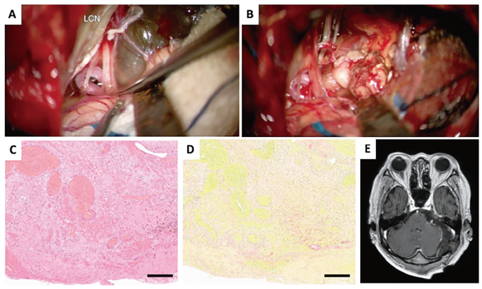
Fig. 4 A: Microscopic image of tumor removal. The cystic lesion was attached to the facial nerve and brainstem in the cerebral cistern. B: Inside the cyst, there was an old hemorrhagic xanthochromic content and a nodule-like lesion resembling a cavernous malformation. C: Original magnification is ×200. Hematoxylin and eosin stain illustrating a mixture of VS and cavernous malformation-like histology, which contains a spindle-shaped cell population consisting of elongated nuclei and eosinophilic endoplasmic reticulum, with a hemorrhage and hemosiderin-phagocytic histiocyte cluster, an inflammatory cell infiltrate consisting mainly of lymphocytes and plasma cells, and a spongiform vascular malformation-like component with numerous small and large dilated thin-walled, irregular muscular vessels. D: Elastica van Gieson stain demonstrated lack of elastic fibers surrounding the vessels. E: More than 95% of the cystic lesion was removed, and ataxia and cranial nerve disturbances improved. Black bar = 250 μm.