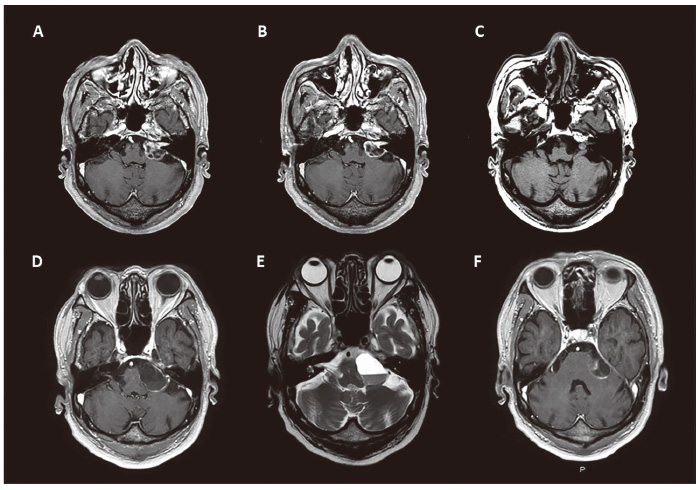
Fig. 3 A: Follow-up image at 3 months after GKS showing transient expansion (tumor volume 4.220 cc). B: Tumor volume showed a decrease from the initial volume of 2.220 cc at 6 months after GKS. C: The tumor volume remarkably decreased to 0.295 cc at 5 years after GKS. D-F: 5 years and 7 months after treatment, the patient developed worsening left hearing loss and ataxic gait, and MRI revealed a large cystic lesion (tumor diameter, 30 mm) with an internal nodule in the left CPA, presenting compression of the brainstem. D, F: axial T1-weighted post-contrast MRI, E: axial T2-weighted MRI.