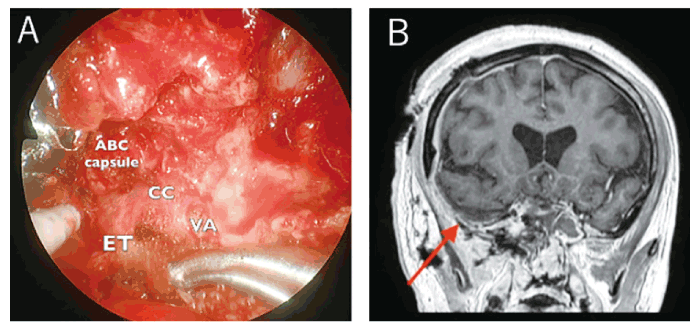
Fig. 3 A; Operative view of resecting ABC capsule in the right infratemporal fossa CC; carotid canal, ET; eustachian tube, VA; vidian artery. B; MRI with contrast enhancement after the secondary surgery. There is no recurrence of the ABCs in the temporal fossa (arrow).